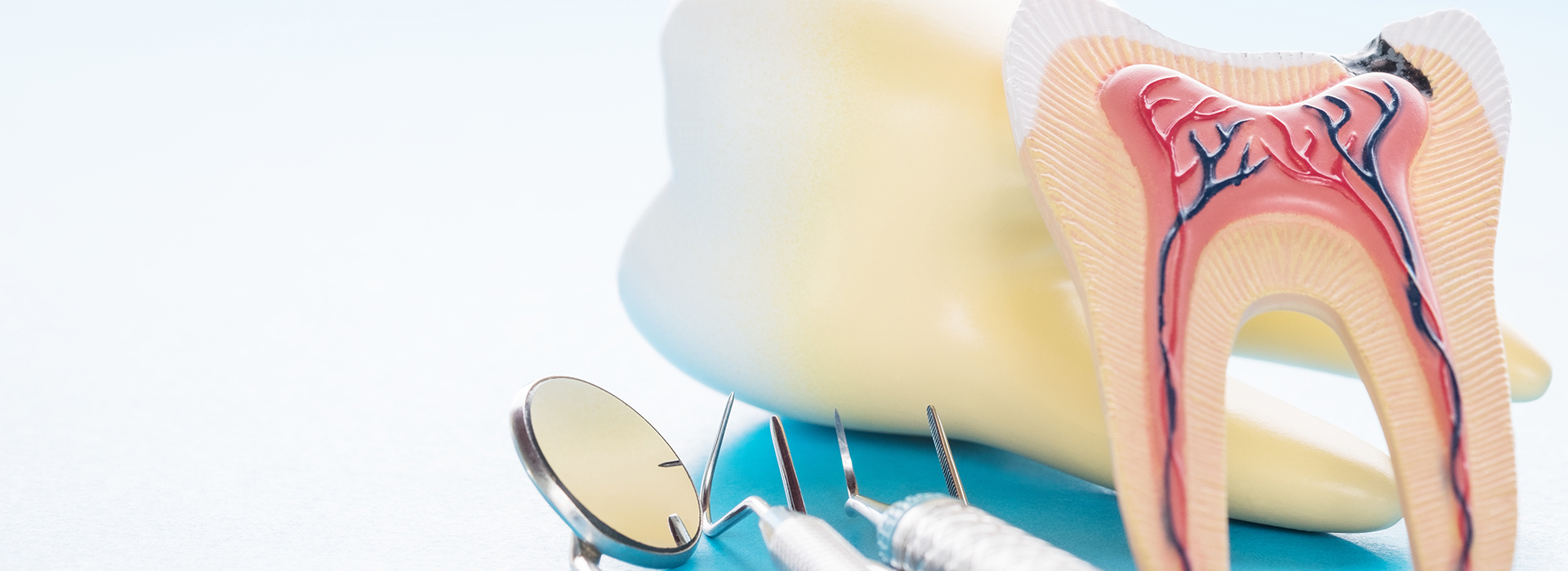
The term “periodontics” refers to the dental specialty that pertains to the prevention, diagnosis and treatment of periodontal disease that affects the gums and jawbone. The gum tissues serve to surround and support the teeth and the underlying jawbone anchors teeth firmly in place. Periodontists have completed several years of extra dental training and are concerned with maintaining the function, health and aesthetics of the jawbone and tissues.
Periodontal disease is a progressive condition which begins with mild gum inflammation called gingivitis. It is the leading cause of tooth loss in adults living in the developed world, and should be taken very seriously. Periodontal disease (often called gum disease) is typically signified by red, swollen, painful, or bleeding gums, but in some cases has no noticeable symptoms.
Periodontal disease generally begins when the bacteria living in plaque cause an infection in the surrounding tissues of the teeth, causing them to become irritated and painful. Eventually, this infection will; cause the jawbone to recede and the tooth to become loose.
In the case of mild/moderate periodontal problems, the focus of the periodontist will be on curing the underlying bacterial infection and then providing advice on the most appropriate home cleaning methods.
Sometimes a deep scaling is needed to remove the bacterial plaque and calculus (tartar) from the teeth and tissues. Where periodontal disease is advanced and the jawbone has regressed significantly, more intensive cleaning may be recommended and loose teeth that cannot be saved will be removed.
The periodontist is trained in all aspects of dental implant procedures, which can restore functionality to the mouth when teeth have been affected by periodontitis.
Because periodontal disease is progressive, it is essential to remove the bacteria and calculus build up to halt the spread of the infection. Your dentist will be happy to advise you on effective cleaning methods and treatment options.
A periodontist is a dentist specializing in the prevention, diagnosis and treatment of infections and diseases in the soft tissues surrounding the teeth, and the jawbone to which the teeth are anchored. Periodontists have to train an additional three years beyond the four years of regular dental school, and are familiar with the most advanced techniques necessary to treat periodontal disease and place dental implants. Periodontists also perform a vast range of cosmetic procedures to enhance the smile to its fullest extent.
Periodontal disease begins when the toxins found in plaque start to attack the soft or gingival tissue surrounding the teeth. This bacterium embeds itself in the gum and rapidly breeds, causing a bacterial infection. As the infection progresses, it starts to burrow deeper into the tissue causing inflammation or irritation between the teeth and gums. The response of the body is to destroy the infected tissue, which is why the gums appear to recede. The resulting pockets between the teeth deepen and if no treatment is sought, the tissue which makes up the jawbone also recedes causing unstable teeth and tooth loss.
There are several ways treatment from a periodontist may be sought. In the course of a regular dental check up, if the general dentist or hygienist finds symptoms of gingivitis or rapidly progressing periodontal disease, a consultation with a periodontist may be recommended. However, a referral is not necessary for a periodontal consultation.
If you experience any of these signs and symptoms, it is important that you schedule an appointment with a periodontist without delay:
Before initiating any dental treatment, the periodontist must extensively examine the gums, jawbone and general condition of the teeth. When gingivitis or periodontal disease is officially diagnosed, the periodontist has a number of surgical and non surgical options available to treat the underlying infection, halt the recession of the soft tissue, and restructure or replace teeth which may be missing.
Periodontal disease is a progressive condition which leads to severe inflammation and tooth loss if left untreated. Antibiotic treatments can be used in combination with scaling and root planning, curettage, surgery or as a stand-alone treatment to help reduce bacteria before and/or after many common periodontal procedures.
Antibiotic treatments come in several different types, including oral forms and topical gels which are applied directly into the gum pockets. Research has shown that in the case of acute periodontal infection, refractory periodontal disease, prepubertal periodontal disease and juvenile periodontal disease, antibiotic treatments have been incredibly effective.
Antibiotics can be prescribed at a low dose for longer term use, or as a short term medication to deter bacteria from re-colonizing.
Oral antibiotics tend to affect the whole body and are less commonly prescribed than topical gel. Here are some specific details about several different types of oral antibiotics:
The biggest advantage of the direct delivery of antibiotics to the surfaces of the gums is that the whole body is not affected. Topical gels and direct delivery methods tend to be preferred over their oral counterparts and are extremely effective when used after scaling and root planing procedures. Here are some of the most commonly used direct delivery antibiotics:
Noticeable periodontal improvements are usually seen after systemic or oral antibiotic treatment. Your Periodontist or dentist will incorporate and recommend the necessary antibiotic treatments as necessary for the healing of your periodontal condition.
Periodontal disease is the leading cause of bone loss in the oral cavity, though there are others such as ill-fitting dentures and facial trauma. The bone grafting procedure is an excellent way to replace lost bone tissue and encourage natural bone growth. Bone grafting is a versatile and predictable procedure which fulfills a wide variety of functions.
A bone graft may be required to create a stable base for dental implant placement, to halt the progression of gum disease or to make the smile appear more aesthetically pleasing.
There are several types of dental bone grafts. The following are the most common:
Dental implants – Implants are the preferred replacement method for missing teeth because they restore full functionality to the mouth; however, implants need to be firmly anchored to the jawbone to be effective. If the jawbone lacks the necessary quality or quantity of bone, bone grafting can strengthen and thicken the implant site.
Sinus lift – A sinus lift entails elevating the sinus membrane and grafting bone onto the sinus floor so that implants can be securely placed.
Ridge augmentation – Ridges in the bone can occur due to trauma, injury, birth defects or severe periodontal disease. The bone graft is used to fill in the ridge and make the jawbone a uniform shape.
Nerve repositioning – If the inferior alveolar nerve requires movement to allow for the placement of implants, a bone grafting procedure may be required. The inferior alveolar nerve allows feeling and sensation in the lower chin and lip.
Bone grafting is a fairly simple procedure which may be performed under local anesthetic; however if large amounts of bone area need to be grafted, general anesthetic may be required.
Initially, the grafting material needs to either be harvested or prepared for insertion. A small incision is made in the gum tissue and then gently separated from the bone. The bone grafting material is then placed at the affected site.
The gum is sutured in place and a follow up appointment will need to be made within 10 days to assess progress. Bone grafting is a highly successful treatment and a good base for further periodontal restorations.
Crown lengthening is generally performed in order to improve the health of the gum tissue, or to prepare the mouth for restorative or cosmetic procedures. In addition, crown lengthening procedures can also be used to correct a “gummy” smile, where teeth are covered with excess gum tissue. Crown lengthening exposes more of the natural tooth by reshaping or recontouring bone and gum tissue. This treatment can be performed on a single tooth, many teeth or the entire gum line, to expose a pleasant, aesthetically pleasing smile.
Crown lengthening is a versatile and common procedure that has many effective uses and benefits. The vast majority of patients who have undergone this type of surgery are highly delighted with the results.
Here are some of the most common reasons for crown lengthening:
Crown lengthening is normally performed under local anesthetic. The amount of time this procedure takes will largely depend in how many teeth are involved and whether a small amount of bone needs to be removed, in addition to the soft tissue. Any existing dental crowns will be removed prior to the procedure, and replaced immediately afterwards.
The dentist will make a series of small incisions around the soft tissue in order to separate the gums away from the teeth. Even if only one tooth requires the re-contour, neighboring teeth are usually treated to provide a more even reshaping. Separating the gums provides the dentist with access to the roots of the teeth and the underlying bone.
In some cases, the removal of a small amount of tissue will provide enough tooth exposure to place a crown. In other cases, the dentist will also need to remove a small amount of bone from around the teeth. The bone is usually removed using a combination of special hand instruments, and rotary instruments. The rotary instruments roughly resemble the drill that is used in cavity treatment.
When the dentist is satisfied the teeth have sufficient exposure, the wound will be cleaned with sterile water and the gum tissue will be sutured with small stitches. The teeth will look noticeably longer immediately after surgery because the gums have now been re-positioned.
The dentist will secure the surgical site using an intraoral (periodontal) bandage, which serves to prevent infection. Prescriptions may be provided for pain medication, and a chlorhexidine (antimicrobial) mouth rinse may be given to help reduce any bacteria attempting to re-colonize. The surgical site will be completely healed in approximately two to three months.
There are many reasons why the gums and jawbone may require corrective treatment, including periodontal disease, trauma and birth defects. Periodontal disease particularly, can greatly disfigure the natural appearance of the gums and teeth and give the smile an unaesthetic appearance.
New “cosmetic surgery” procedures are now available in periodontics, which effectively correct cosmetic problems and restore natural beauty to the smile.
Here are some of the most common gum and jawbone corrective treatments:
A gum graft (also known as a gingival graft or periodontal plastic surgery), is a collective name for surgical periodontal procedures that aim to cover an exposed tooth root surface with grafted oral tissue.
Exposed tooth roots are usually the result of gingival recession due to periodontal disease. There are other common causes, including overly aggressive brushing and trauma
Gum grafting is a common periodontal procedure. Though the name might sound frightening, the procedure is commonly performed with excellent results.
Once the need for gum grafting surgery has been determined, there are several treatments the dentist will want perform before gum grafting takes place. First, the teeth must be thoroughly cleaned supra and subgingivally to remove calculus (tartar) and bacteria. The dentist can also provide literature, advice and educational tools to increase the effectiveness of homecare and help reduce the susceptibility of periodontal disease in the future.
The gum grafting procedure is usually performed under local anesthetic. The exact procedure will depend much on whether tissue is coming from the patient’s palate or a tissue bank.
Initially, small incisions will be made at the recipient site to create a small pocket to accommodate the graft. Then a split thickness incision is made and the connective tissue graft is inserted into the space between the two sections of tissue. The graft is usually slightly larger than the recession area, so some excess will be apparent.
Sutures are often placed to further stabilize the graft and to prevent any shifting from the designated site. Surgical material is used to protect the surgical area during the first week of healing. Uniformity and healing of the gums will be achieved in approximately six weeks.
Gingival recession (receding gums) refers to the progressive loss of gum tissue, which can eventually result in tooth root exposure if left untreated. Gum recession is most common in adults over the age of 40, but the process can begin in the teenage years.
Gum recession can be difficult to self-diagnose in its earlier stages because the changes often occur asymptomatically and gradually. Regular dental check ups will help to prevent gum recession and assess risk factors.
The following symptoms may be indicative of gum recession:
Gum recession is an incredibly widespread problem that dentists diagnose and treat on a daily basis. It is important to thoroughly examine the affected areas and make an accurate diagnosis of the actual underlying problem. Once the cause of the gum recession has been determined, surgical and non surgical procedures can be performed to halt the progress of the recession, and prevent it from occurring in the future.
Every case of gum recession is slightly different, and therefore many treatments are available. The nature of the problem which caused the recession to begin with needs to be addressed first.
If overly aggressive brushing techniques are eroding the gums, a softer toothbrush and a gentler brushing technique should be used. If poor oral hygiene is a problem, prophylaxis (professional dental cleaning) may be recommended to rid the gum pockets of debris and bacteria. In the case of a severe calculus (tartar) build up, scaling and root planing will be performed to heal the gingival inflammation and clean the teeth.
Once the cause of the gingival recession has been addressed, surgery of a more cosmetic or restorative nature may be recommended. Gum tissue regeneration and gum grafting are two excellent ways to restore natural symmetry to the gums and make the smile look more aesthetically pleasing.
Periodontal disease is a progressive condition which leads to gum inflammation, the recession of bone and gum tissue, and tooth loss if left untreated. There are a variety of effective treatments and procedures available, including pocket irrigation, which can assist in treating the progression of the disease.
Pocket irrigation aims to cleanse plaque from the interdental (between teeth) and subgingival (under the gumline) regions of the mouth in order to prevent the colonization of harmful oral bacteria. This procedure is also used to deliver antibacterials to the subgingival areas.
Pocket irrigation, which is also known as oral irrigation, is a versatile dental treatment used for several different preventative purposes. Pocket irrigation may be performed as part of a professional dental cleaning, or at home with a specially modified oral irrigator.
Pocket irrigation is generally performed in combination with other dental treatments; for example, as part of a professional dental cleaning or in combination with pocket reduction surgery.
In the case of a pocket reduction surgery, the tartar and subgingival plaque will be removed with special scaling and root planing instruments. When the pockets are free of debris, an antimicrobial agent may be applied using an oral irrigator. This will help reduce harmful oral bacteria which still remain in the pockets.
During a deep cleaning procedure, a pocket irrigator may be used after scaling and root planing to cleanse the pockets. Again, an antimicrobial agent may be applied to help reduce subgingival oral bacteria.
Pocket irrigation can be performed at home as part of the daily oral routine using a water jet or water pick. Flushing the interdental area with water is less harmful to soft tissue than dental floss, but should not be used as a substitute for professional dental cleaning.
Pocket reduction surgery (also known as gingivectomy, osseous surgery and flap surgery) is a collective term for a series of several different surgeries aimed at gaining access to the roots of the teeth in order to remove bacteria and tartar (calculus).
The human mouth contains dozens of different bacteria at any given time. The bacteria found in plaque (the sticky substance on teeth) produce acids that lead to demineralization of the tooth surface, and ultimately contribute to periodontal disease.
Periodontal infections cause a chronic inflammatory response in the body that literally destroys bone and gum tissues once they invade the subgingival area (below the gum line). Gum pockets form and deepen between the gums and teeth as the tissue continues to be destroyed.
Periodontal disease is a progressive condition which, if left untreated, causes massive bacteria colonization in gum pockets can eventually lead to teeth falling out. Pocket reduction surgery is an attempt to alleviate this destructive cycle, and reduce the depth of the bacteria-harboring pockets.
Pocket reduction surgery is a common periodontal procedure which has been proven effective at eliminating bacteria, reducing inflammation and saving teeth. The goals of pocket reduction surgery are:
Before recommending treatment or performing any procedure, the dentist will perform thorough visual and x-ray examinations in order to assess the condition of the teeth, gums and underlying bone. Pocket reduction surgery may be performed under local or general anesthetic depending on the preferences of the patient.
The gums will be gently pulled back from the teeth and bacteria and calculus (tartar) will be eliminated. Scaling and root planing will generally be required to fully remove the ossification (tartar) from the surface of the tooth root. If the root is not completely smooth, a planing procedure will be performed to ensure that when the gums do heal, they will not reattach to rough or uneven surfaces.
The final part of the surgery is usually the administration of an antimicrobial liquid to eliminate any remaining bacteria and promote healing. The gum is then sutured with tiny stitches that are left in place for 5-10 days.
Though the gums will be more sensitive immediately following the procedure, there will be a significant reduction in pocket depth and a vast improvement in the condition of the teeth and gums.
Periodontal disease is an extremely serious progressive condition which can lead to tooth loss if left untreated. Periodontal disease occurs when a bacterial infection in the gum pockets causes the bone and gum tissue to recede away from the teeth. The quality and quantity of bone and gum tissue is greatly reduced as the gum pockets continue to deepen. Fortunately, prompt diagnosis combined with effective regenerative procedures can halt the progression of the disease and save the teeth.
There are many regenerative dental procedures available to restore health to the gums, bone and teeth. The following are the most commonly used regenerative procedures:
Dental implants and other restorative procedures depend on the jawbone being sufficiently strong and thick. When periodontal disease has taken hold, the jawbone may have become too thin to successfully support an implant. Natural bone regeneration (or bone grafting) is one of the best ways to promote growth and thicken the jawbone.
Initially, bone grafting material is obtained from elsewhere on the body or from a bone bank. The gums are gently pulled back, and an opening is made in the jawbone where the grafting material will be packed in tightly. A membrane is used to maintain the shape of the new bone, and tissue stimulating proteins and a platelet-rich growth factor may be applied to promote faster healing. The surgical site will be sutured and packed with protective material. The bone will naturally regenerate and become strong enough to eventually support an implant.
Gum tissue regeneration is usually performed by way of gum grafting. When gum tissue has been lost as a result of periodontal disease, the smile may appear discolored, toothy or unpleasant. A gum graft aims to symmetrically contour the gum line, cover any exposed tooth roots and restore health to the gums. Initially, a small strip of tissue will be removed from the roof of the mouth and placed in the middle of a split-thickness incision at the recipient site. The surgical site will be covered with a protective membrane which promotes tissue regeneration, covered in platelet-rich growth factor and tissue stimulating proteins, and then sutured closed. The soft tissue surrounding the teeth will naturally regenerate, producing a healthy, smile.
The aim of pocket reduction surgery is to reduce the depth of the gum pockets and make it much harder for bacteria to invade the gum tissue. Initially, the gum tissue is pulled back from the teeth and the subgingival area is thoroughly cleansed under local anesthesia. Extensive scraping may be required to fully remove the plaque, debris and tartar from below the gum line. An antimicrobial agent may then be applied to the affected area to kill any remaining bacteria, and the surgical site will be sutured closed. Pocket reduction surgery is very effective for halting tissue loss and promoting the natural healing of the gums.
The term “scaling” refers to the removal of plaque and calculus (tartar) from the supragingival region (above the gum line). This treatment is usually conducted in the course of a routine professional cleaning, and performed under local anesthetic. Root planing usually accompanies scaling and is performed in the subgingival region (below the gum line). Tartar and toxins are initially removed, and then the tooth root is smoothed down to promote good healing in the gum tissue. The planing of the tooth root provides a smooth area which prevents bacteria from recolonizing. If these procedures do not completely eradicate the infection, surgical measures may be required.
Ridge modification is an effective procedure for treating deformities in the upper and lower jaws. These deformities can occur as a result of periodontal disease, trauma, injury, wearing dentures, or developmental problems. Such defects can leave insufficient bone for the placement of dental implants and an additional unattractive indentation in the jaw line adjacent to the missing teeth.
During the ridge modification procedure, the gum is lifted away from the ridge to fully expose the defect in the bone. The bony defect can be filled with bone graft material which can help regenerate lost bone, or a bone substitute. Finally, the incision is closed and several months of healing will be required. Depending on the case and type of implant and procedure, a dental implant may be placed during the ridge modification procedure or when healing is complete; much depends on the precise condition of the bone. Ridge modification improves the cosmetic appearance, functionality of the mouth, and the chance of enjoying dental implants for many years.
Root amputation is a specialized dental procedure, whereby one root is removed from a multi-root tooth. The tooth is then stabilized and rendered fully functional with a crown or filling. The multi-root teeth best suited to the root amputation procedure are the molars at the back of the mouth. These large flat teeth have either two or three roots depending on whether they are situated on the upper or lower jaw.
The general purpose of root amputation is to save an injured or diseased tooth from extraction. Most dentists agree that there is no better alternative than retaining a healthy natural tooth, and the root amputation procedure makes this possible. Dental implants, extensive bridgework and custom-made tooth replacements can be expensive and time-consuming. Generally, root amputation and the necessary crown work are less expensive and can be completed in 1-3 short visits.
It is important to note that root amputation can only be performed on an otherwise healthy tooth. Even in the case of a “key” tooth, extraction will be performed if the tooth is diseased, badly fractured or otherwise injured. Suitable teeth for root amputation have a healthy tooth surface, strong bone support and healthy underlying gums.
Prior to root amputation, it is necessary to perform root canal treatment. The amputation itself involves cutting deep into the tooth where blood vessels and nerves are located. For this reason, the pulp of the tooth including these vessels and nerves needs to be removed before resectioning the roots. The root canal and amputation treatments will be performed under local anesthetic.
During the root amputation procedure, a small incision will be created in the gum to fully expose the roots of the affected tooth. The root will be sectioned off from the rest of the tooth and then removed. To kill any remaining bacteria, the whole area will be cleansed with saline solution, and then sutures (stitches) will be applied to seal the incision.
Finally, a temporary crown or filling will be placed to secure the tooth. Depending on the specific situation, painkillers, antibiotics and a medicated anti-microbial mouthwash may be prescribed. In 7-10 days, the stitches will be removed and the gum will have healed. Arrangements can now be made to place the permanent crown or filling.
A dental implant is essentially an artificial tooth root which is attached to the jaw bone. Eventually, a replacement tooth or bridge will be firmly fixed to this root, restoring complete function to the tooth. The key to a successful and long-lasting implant is the quality and quantity of jawbone to which the implant will be attached. If bone has been lost due to injury or periodontal disease, a sinus augmentation can raise the sinus floor to allow for new bone formation.
In the most common sinus augmentation technique, a tiny incision is made near the upper premolar or molar region to expose the jawbone. A small opening is cut into the bone and the membrane lining the sinus on the other side of the opening is gently pushed upward. The underlying space is filled with bone graft material and the incision is closed. The bone which is used for this procedure may be from your own body or from a cadaver. Sometimes the dentist might use synthetic materials which can also stimulate bone formation. The implants are placed after healing has occurred; this will depend on the individual case. Sinus augmentation has been shown to increase the success of dental implant procedures.
Soft tissue grafting is often necessary to combat gum recession. Periodontal disease, trauma, aging, over brushing, and poor tooth positioning are the leading causes of gum recession which can lead to tooth-root exposure in severe cases.
When the roots of the teeth become exposed, eating hot and cold foods can be uncomfortable, decay is more prevalent and the aesthetic appearance of the smile is altered. The main goal of soft tissue grafting is to either cover the exposed root or to thicken the existing gum tissue in order to halt further tissue loss.
Soft tissue grafting is an extremely versatile procedure that has many uses. Recent developments in dental technology have made soft tissue grafting more predictable and less intrusive. Here are some of the main benefits associated with soft tissue grafting treatment:
Initially, deep cleaning will be performed both above and below the gum line to clear the teeth and roots of calculus (tartar). The grafting procedure itself will generally be performed under local anesthetic, but this will depend on the size of the areas receiving grafts. A small incision will be made at the recipient site in order to create a small pocket. A split thickness incision is made in this pocket and the donor tissue is placed between the two sections of this area. The donor tissue strip is generally larger than the incision, so some excess will be apparent.
Platelet rich growth factors which stimulate natural tissue growth and promote good healing may be applied to the site before suturing. In addition, tissue-stimulating proteins may be added to encourage quicker tissue growth. Finally, the wound site will be sutured to prevent shifting, and surgical material will be placed to protect the sensitive area. Gum uniformity and substantial healing will take place in the first six weeks after the procedure.
